Lumbar Pain Treatment by Radiofrequency
A safe and effective minimally invasive technique to treat pain related to facet joint osteoarthritis. Performed under precise radiological guidance by an expert team.
What is the principle of radiofrequency?
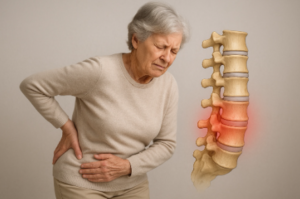
This is a technique used in interventional radiology with several applications. Here, the goal is to relieve one cause of low back pain: facet joint osteoarthritis.
Pain related to this osteoarthritis is transmitted to the brain via small nerves. Schematically, a nerve can be compared to an electrical cable with wires inside and an outer sheath. Radiofrequency damages the cable sheath, preventing the pain signal from reaching the brain.
Does radiofrequency treat osteoarthritis?
No. Radiofrequency treats pain secondary to osteoarthritis, but the main cause, osteoarthritis, continues to progress.
Osteoarthritis is a physiological phenomenon, meaning normal from a certain age depending on several factors (obesity, occupational life, genetics, etc.), and not all osteoarthritis is painful.
Radiofrequency is therefore part of the therapeutic arsenal for facet joint osteoarthritis.
What examinations should be performed before radiofrequency?
It is preferable to have had a prior consultation with your general practitioner or a specialist, as well as imaging workup including MRI or bone scan. Standard X-rays are no longer sufficient today to precisely determine the various causes of low back pain.
There is then the diagnostic block step, which consists of injecting a dose of anesthetic under guidance along the nerve pathways to temporarily "numb" them. If pain decreases by 50% after the block, the test is considered positive and radiofrequency can be offered.
How is radiofrequency performed?
Radiofrequency is an outpatient procedure with an estimated duration of approximately 30 minutes and anesthetic sedation for patient comfort.
- The patient lies on their stomach. An intravenous catheter is placed for anesthesia and sedation, along with adhesive pads on the thighs for the procedure.
- The lower back is disinfected before starting the procedure with local anesthesia in addition to anesthetic sedation for maximum comfort.
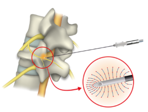
- Radiofrequency needles are placed under fluoroscopic guidance. The number of needles varies depending on the procedure and clinical scenario.
- Once in place, a preliminary motor and sensory test is performed to ensure needles are correctly positioned, particularly away from motor spinal nerves.
- Heating through the needles can then begin and generally lasts 60 seconds per needle.
- The needles are then removed and simple dressings are applied before return to the outpatient unit where the patient is generally kept for 2 hours before discharge home.
Discharge home must be accompanied given the anesthesia. A transport voucher can be arranged for isolated patients.
What is the recovery like? Will I be relieved immediately?
Immediate recovery is marked by transient pain along the needle tracts for 24–48 hours.
Return to activities should be gradual from 48 hours onward and during the following week: avoid unusual physical exertion during the first 7 days.
Pain relief generally occurs within 6 weeks following the procedure. This delay must be waited before judging effectiveness.
As a reminder, the goal is a 50% reduction in painful symptoms, which remains subjective. Complete disappearance may occur in some cases but is not the rule.
Is it a permanent treatment?
There is a risk of recurrence because the procedure damages the nerve myelin sheath without completely destroying the nerve. This sheath will regenerate and pain may recur, although this occurs after several months or even a year.
If relief was significant over a long period, the procedure can be repeated.
What are the possible complications?
The risks associated with this procedure are very low but exist, as with all interventions.
Risks common to all procedures:
- Hemorrhagic risk: hematoma along the tract. However, the caliber of needles used is small, minimizing this risk. It is important to report antiplatelet or anticoagulant medication during consultation.
- Infectious risk: also low because there is no skin opening and the procedure is performed in an interventional radiology suite under aseptic conditions.
- Anesthesia-related risks: allergy to products used during anesthesia or decompensation of underlying pathology (cardiovascular, respiratory, etc.).
Risks specific to analgesic radiofrequency:
- Tingling in the legs: related to nerve irritation secondary to the procedure. Most often spontaneously regressive within a few days.
- Motor deficit in the leg: serious theoretical complication related to incorrect needle positioning. This exceptional complication is prevented by the motor test performed once the needle is in place and before heating begins, as well as by precise imaging means to control needle positioning.
Are there contraindications?
The main contraindications to these procedures are related to the presence of the following devices:
- Pacemaker
- Defibrillator
- Neurostimulation device
Pregnancy is of course a contraindication due to the use of X-rays.
What are the alternatives?
Radiofrequency treatment is a second-line treatment. It is generally indicated after first-line treatment consisting of medication and physiotherapy.
Facet joint injections under CT or fluoroscopic guidance are then offered. They can relieve pain but their duration of effectiveness over time is limited.
Analgesic radiofrequency follows, with a longer-lasting effect over time.
In case of failure or early recurrence of low back pain related to facet joint osteoarthritis, the surgical option remains: fixation of the affected vertebral segment (spinal fusion).
Ready to book an appointment?
Contact us to discuss your situation and schedule your consultation
Book an appointment